Chest Muscles Diagram : Muscles Of The Pectoral Region Major Minor Teachmeanatomy. The chest muscles of our body can get pulled and strained. Muscle chart of the groin 12 photos of the muscle chart of the groin muscle chart groin, muscles of the groin area diagram, human muscles, muscle chart groin, muscles of the groin area diagram. The muscles of the chest and upper back occupy the thoracic region of the body inferior to the neck and superior to the abdominal region and include the muscles of the shoulders. Muscle of the forearm quiz The intercostal muscles form the chest wall and function in respiration.
A muscle strain or pull happens when your muscle is stretched or torn. The terms pulled muscle and muscle strain refer to an injury that involves an overstretched or torn muscle. Human body muscle system, the muscles of the human body that work the skeletal system, that are under voluntary control, and that are concerned with movement, posture, and balance. The major muscle in the chest is the pectoralis major. Related posts of chest muscles diagram elbow muscle anatomy mri.
Easy Notes On The Pectoral Region Muscles Learn In Just 6 Mins Earth S Lab from www.earthslab.com Chest wall pain is caused by problems affecting the muscles, bones and/or nerves of the chest wall. The chest, as part of this group, enables you to perform pushing actions such as the barbell bench press or a daily activity such as moving a heavy dresser. The dominant muscle in the upper chest is the pectoralis major. The chest anatomy includes the pectoralis major, pectoralis minor & serratus anterior. Anatomy muscle function 12 photos of the anatomy muscle function anatomy muscle function, anatomy muscle function quiz, function of muscle anatomy, human muscle anatomy function, muscle anatomy and function, human muscles, anatomy muscle function, anatomy muscle function quiz, function of muscle anatomy, human. The intercostal muscles form the chest wall and function in respiration. Doctors diagnose chest wall pain in at least 25% of patients who come to the emergency room for chest pain. The obliques are abdominal muscles that assist during bending and twisting of the torso.
The chest is the area of origin for many of the body's systems as it houses organs such as the heart, esophagus, trachea, lungs, and thoracic diaphragm.
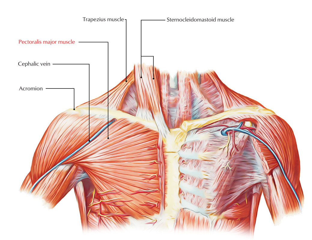
This is because er doctors usually are focused on. The pectoralis major, pectoralis minor, serratus anterior and subclavius. A person with a muscle strain in the chest may experience sudden, sharp pain in this area. The dominant muscle in the upper chest is the pectoralis major. The pectoral region is located on the anterior chest wall. The chest, as part of this group, enables you to perform pushing actions such as the barbell bench press or a daily activity such as moving a heavy dresser. The major muscle in the chest is the pectoralis major. Chest wall pain is caused by problems affecting the muscles, bones and/or nerves of the chest wall. Function of the chest muscles. Be sure to visit the guide for more context and information about muscles of the chest diagram, or read some of our other health. Chest muscles diagram, picture of chest muscles diagram. Muscle chart of the groin 12 photos of the muscle chart of the groin muscle chart groin, muscles of the groin area diagram, human muscles, muscle chart groin, muscles of the groin area diagram. In this image, you will find frontalis, orbicularis oculi, zygomaticus, masseter, orbicularis oris, sternocleidomasteoid, deltoid, pectoralis major, biceps brachii, iliopsoas, adductor longus, gastrocnemius.
The chest is the area of origin for many of the body's systems as it houses organs such as the heart, esophagus, trachea, lungs, and thoracic diaphragm. The muscle is strong enough to pump up to 2,000 gallons — as much as a fire department's tanker truck — of blood through one's body every day. The terms pulled muscle and muscle strain refer to an injury that involves an overstretched or torn muscle. Muscle of the forearm quiz Unfortunately, in many cases, that's as far as the doctor takes the diagnosis.
Human Chest Muscles Pectoralis Major Illustration Stock Photo Alamy from c8.alamy.com The circulatory system does most of its. Muscle anatomy practice exam 12 photos of the muscle anatomy practice exam anatomy muscle practice questions, muscle anatomy practice exam, human muscles, anatomy muscle practice questions, muscle anatomy practice exam Click the answer to find similar crossword clues. Muscle chart of the groin 12 photos of the muscle chart of the groin muscle chart groin, muscles of the groin area diagram, human muscles, muscle chart groin, muscles of the groin area diagram. Related posts of chest muscle diagram muscle anatomy upper limb. A muscle strain or pull happens when your muscle is stretched or torn. Be sure to visit the guide for more context and information about muscles of the chest diagram, or read some of our other health. In this image, you will find frontalis, orbicularis oculi, zygomaticus, masseter, orbicularis oris, sternocleidomasteoid, deltoid, pectoralis major, biceps brachii, iliopsoas, adductor longus, gastrocnemius.
Human body muscle system, the muscles of the human body that work the skeletal system, that are under voluntary control, and that are concerned with movement, posture, and balance.
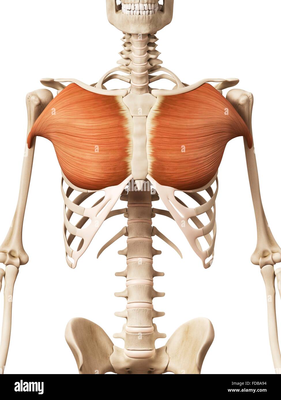
Muscle chart of the groin 12 photos of the muscle chart of the groin muscle chart groin, muscles of the groin area diagram, human muscles, muscle chart groin, muscles of the groin area diagram. The terms pulled muscle and muscle strain refer to an injury that involves an overstretched or torn muscle. The dominant muscle in the upper chest is the pectoralis major. Your pectoralis major and pectoralis minor muscles make up most of the muscle mass in your chest. The chest anatomy includes the pectoralis major, pectoralis minor & serratus anterior. Enter the answer length or the answer pattern to get better results. Related posts of muscle diagram for chest and back anatomy muscle function. See chest anatomy stock video clips. The major muscle in the chest is the pectoralis major. This chest press variation allows you to work different angles of the pectoral muscles and assists with overall chest strengthening. The obliques are abdominal muscles that assist during bending and twisting of the torso. The chest is part of a larger group of pushing muscles found in the upper body. Muscles allow a person to move, speak, and chew.
Function of the chest muscles. The major muscle in the chest is the pectoralis major. Muscle of the forearm quiz If you are pulling your muscles more than enough and doing burdening lots of effort, then the pain that is going to felt by you is no the symptom of the muscular chest pain. The circulatory system does most of its.
Amazon Com Male Chest Muscles Labeled Educational Medical Chart Black Wood Framed Art Poster 20x14 Home Kitchen from images-na.ssl-images-amazon.com Set a bench to a 45 degree incline. Elbow muscle anatomy mri 12 photos of the elbow muscle anatomy mri elbow muscle anatomy axial, elbow muscle anatomy mri, human muscles, elbow muscle anatomy axial, elbow muscle anatomy mri The chest, as part of this group, enables you to perform pushing actions such as the barbell bench press or a daily activity such as moving a heavy dresser. The circulatory system does most of its. Create bulk of the chest, commonly known as pecs sternoicleidomastoid connects from the mastoid process at the back/base of head to the sternum and clavicle The pectoral region is located on the anterior chest wall. Your pectoralis major and pectoralis minor muscles make up most of the muscle mass in your chest. The muscle is strong enough to pump up to 2,000 gallons — as much as a fire department's tanker truck — of blood through one's body every day.
Chest muscles diagram, picture of chest muscles diagram.
The pectoralis major and minor. In this image, you will find frontalis, orbicularis oculi, zygomaticus, masseter, orbicularis oris, sternocleidomasteoid, deltoid, pectoralis major, biceps brachii, iliopsoas, adductor longus, gastrocnemius. Create bulk of the chest, commonly known as pecs sternoicleidomastoid connects from the mastoid process at the back/base of head to the sternum and clavicle The obliques are abdominal muscles that assist during bending and twisting of the torso. Function of the chest muscles. If you are pulling your muscles more than enough and doing burdening lots of effort, then the pain that is going to felt by you is no the symptom of the muscular chest pain. Chest muscle in women body. Related posts of chest muscle diagram muscle anatomy upper limb. Chest muscle anatomy diagram : Human body muscle system, the muscles of the human body that work the skeletal system, that are under voluntary control, and that are concerned with movement, posture, and balance. Click the answer to find similar crossword clues. The muscle is strong enough to pump up to 2,000 gallons — as much as a fire department's tanker truck — of blood through one's body every day. The chest, as part of this group, enables you to perform pushing actions such as the barbell bench press or a daily activity such as moving a heavy dresser.